|

|
|
Dear Colleague,
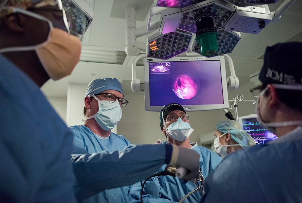
There is much discussion around the pros and cons of both the open-hysterotomy and fetoscopic techniques for prenatal myelomeningocele repair. Which procedure is the best option for expecting patients?
At Texas Children’s Fetal Center, we have found that the outcomes of patients using the fetoscopic approach are comparable to the standard MOMS trial outcomes, with significant added benefits that come from using a less-invasive approach. Although we still offer open-hysterotomy and are experienced with both surgical methods, the exciting results of our experimental fetoscopic approach has fetal centers from around the world coming to us for education on our technique.
Below, we have included some information about the fetoscopic approach and its benefits as compared to both the open-hysterotomy and postnatal repair, details about the procedure, inclusion and exclusion criteria, and outcomes data. Also, we have included a link to a recently published paper authored by our fetal surgeons that highlights the decrease in risk of uterine rupture with the fetoscopic approach.
We hope you will find this information helpful in providing the best recommendations for your patients. Please feel free to contact us with any questions you may have.
Sincerely,
Michael A. Belfort, MBBCH, DA(SA), MD (Cape Town), PhD, FRCSC, FRCOG, FACOG
Obstetrician and Gynecologist-in-Chief
Texas Children’s Hospital
Department of Obstetrics and Gynecology
Baylor College of Medicine
|
|
|
|
|
|
|
BENEFITS TO MOM AND BABY
|
|
Below are the benefits to both mother and baby for this procedure as compared to the
open-hysterotomy and postnatal repair procedures.
Benefits compared to the open-hysterotomy procedure
- Provide an opportunity for a vaginal delivery. This intervention does not require a cesarean delivery like the open-hysterotomy approach. As of January 2019, more than 50 percent of our patients who had a fetoscopic repair have had a vaginal delivery.
- Reduce risk of uterine dehiscence. The very small incisions in the uterus using the fetoscopic approach significantly reduce the risk of uterine thinning or dehiscence on the scar tissue of the sites of access, which contrasts with the 36 percent risk that is present in the open-hysterotomy approach. If vaginal delivery is achieved, complications related to uterine scar thinning or abnormally invasive placenta (percreta, accreta, increta) may be reduced in subsequent pregnancies.
- Avoid risk of early delivery and increase gestation time. Average gestational age at delivery for those who undergo the fetoscopic procedure is 37-38 weeks, as compared to 34 weeks in the MOMS trial. This helps avoid many of the risks to the baby associated with premature birth,
Benefits compared to postnatal repair
- Prevent or reduce shunt rate. The number one goal of the fetoscopic procedure is to decrease the risk of hydrocephalus and/or need for a ventriculoperitoneal shunt to drain excess cerebrospinal fluid postnatally. To date, we’ve reduced the number of patients who need a postnatal shunt by about 50 percent, similar to what is reported by the MOMS trial.
- Preserve nerve function and ambulation. The second goal of the fetoscopic procedure is to preserve as much nerve function as possible by closing the lesion in-utero to protect the spinal nerves from exposure, helping improve the function of the legs and potentially the child’s ability to walk. Results of the fetoscopic method are similar to what is reported by the MOMS trial.
For more detailed information about our outcomes, please reference:
Fetoscopic Open Neural Tube Defect Repair: Development and Refinement of a Two-Port, Carbon Dioxide Insufflation Technique. Obstet Gynecol. 2017 Mar 06. PMID: 28277363. Belfort MA, Whitehead WE, Shamshirsaz AA, Bateni ZH, Olutoye OO, Olutoye OA, Mann DG, Espinoza J, Williams E, Lee TC, Keswani SG, Ayres N, Cassady CI, Mehollin-Ray AR, Sanz Cortes M, Carreras E, Peiro JL, Ruano R, Cass DL.
|
|
|
|
INCLUSION AND EXCLUSION CRITERIA
Comprehensive evaluation of each individual pregnancy is essential to determine whether fetal surgery for myelomeningocele is an appropriate intervention.

OUTCOMES DATA
Texas Children’s Fetal Center has pioneered the fetoscopic repair of spina bifida approach and completed more cases than any other center in the United States.

RESEARCH
At Texas Children’s Fetal Center, we are dedicated to not only offering the best available treatments for our patients, but to finding new and better treatments that offer the best possible outcomes. As a national leader in research, we are conducting an exhaustive study on every aspect of fetoscopic myelomeningocele repair, with results being reported to the scientific community as frequently as possible. Here are some of our most important publications on the procedure to date:
Does fetoscopic or open repair for spina bifida affect fetal and postnatal growth?
Sanz Cortes, Davila I, Torres P, Yepez M, Lee W, Guimaraes CV, Sangi-Haghpeykar H, Whitehead W, Castillo J, Nassr AA, Espinoza J, Shamshirsaz A, Belfort M.
Prenatal brain imaging for predicting postnatal hydrocephalus treatment in fetuses that had neural tube defect repair.
Zarutskie A, Guimaraes C, Yepez M, Torres P, Shetty A, Sangi-Haghpeykar H, Lee W, Espinoza J1, Shamshirsaz A, Nassr A, Belfort M, Whitehead W, Sanz Cortes M.
Amniotic membrane and placental histopathological findings after open and fetoscopic prenatal neural tube defect repair.
Sanz Cortes M, Castro E, Sharhan D, Torres P, Yepez M, Espinoza J, Shamshirsaz AA, Nassr AA,
Popek E, Whitehead W, Belfort MA
Doppler changes in umbilical artery and ductus venosus during fetoscopic prenatal surgical repair of myelomeningocele. Kassir E, Belfort MA, Shamshirsaz AA, Sanz Cortes M, Whitehead WE, Espinoza J.
Management of Labor and Delivery After Fetoscopic Repair of an Open Neural Tube Defect.
Kohn JR, Rao V, Sellner AA, Sharhan D, Espinoza J, Shamshirsaz AA, Whitehead WE, Belfort MA,
Sanz Cortes M.
Fetoscopic Open Neural Tube Defect Repair: Development and Refinement of a Two-Port, Carbon Dioxide Insufflation Technique.
Obstet Gynecol. 2017 Mar 06. PMID: 28277363. Belfort MA, Whitehead WE, Shamshirsaz AA, Bateni ZH, Olutoye OO, Olutoye OA, Mann DG, Espinoza J, Williams E, Lee TC, Keswani SG, Ayres N, Cassady CI, Mehollin-Ray AR, Sanz Cortes M, Carreras E, Peiro JL, Ruano R, Cass DL.
Texas Children’s Spina Bifida Program
Exceptional postnatal care for spina bifida patients at Texas Children’s Hospital
Texas Children’s provides a comprehensive neurodevelopmental and postnatal follow-up program for patients born with spina bifida led by Dr. Jonathan Castillo Porter, developmental pediatrician. For more information about Texas Children’s Spina Bifida Program, please call us with any questions you may have at 832-822-4308.
|
|
|
Contact Texas Children’s Fetal Center
Texas Children’s Fetal Center welcomes all referrals. Candidates are evaluated carefully and accepted for maternal-fetal surgery on a case-by-case basis. Please contact us at
832-822-2229 if you would like to discuss the eligibility of your patient for fetal surgery.
How to refer a patient
To refer a patient, visit our website and fill out our online referral form. Patient historical information and medical records can be uploaded online in the form as well. Please be sure to fill out every field in the form, if possible. Please call us if there are any questions with the online form submission or if there are any urgent matters. Our phones are answered 24 hours a day, 7 days a week.

|
|
|
|
Connect with Texas Children's
Hospital:
|
|
|
|
© 2019 Texas Children's Hospital. All
Rights Reserved.
6621 Fannin Street |
Houston, Texas, 77030
|
|
|
|


