
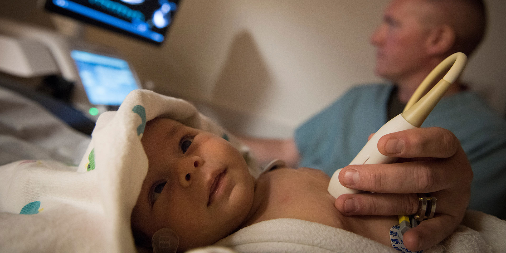
Our noninvasive imaging lab performs more than 34,000 echocardiograms annually for fetuses, infants, children and adults with congenital or acquired heart disease. We provide transesophageal, epicardial, intracardiac and transthoracic echocardiographic support in the catheterization labs, operating rooms and intensive care units on a daily basis. Our imaging team now consists of 38 highly trained sonographers and more than 35 faculty members, a dedicated advanced practice provider for sedation, as well as nurses, medical assistants and clerical staff.
Housed in the Lester and Sue Smith Legacy Tower, we have 15 transthoracic echo imaging rooms with a six-bed sedation bay. For our clinic patients, unique pods nest echo imaging rooms adjacent to clinic rooms to promote communication between imaging and clinical teams and improve our patient/family experience. State-of-the-art multi-physician “Mission Control” reading rooms are embedded between our pods, giving echo faculty the ability to watch live imaging from within the echo lab and in patient care units throughout the hospital.
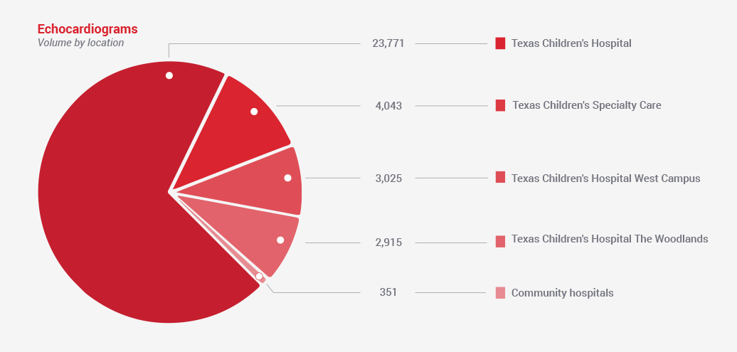
Echocardiographic services are also provided at all of the Texas Children’s Specialty Care locations and we have additional imaging centers at Texas Children’s Hospital West Campus and Texas Children’s Hospital The Woodlands to support our services in the community.
Cardiac MRI & CT
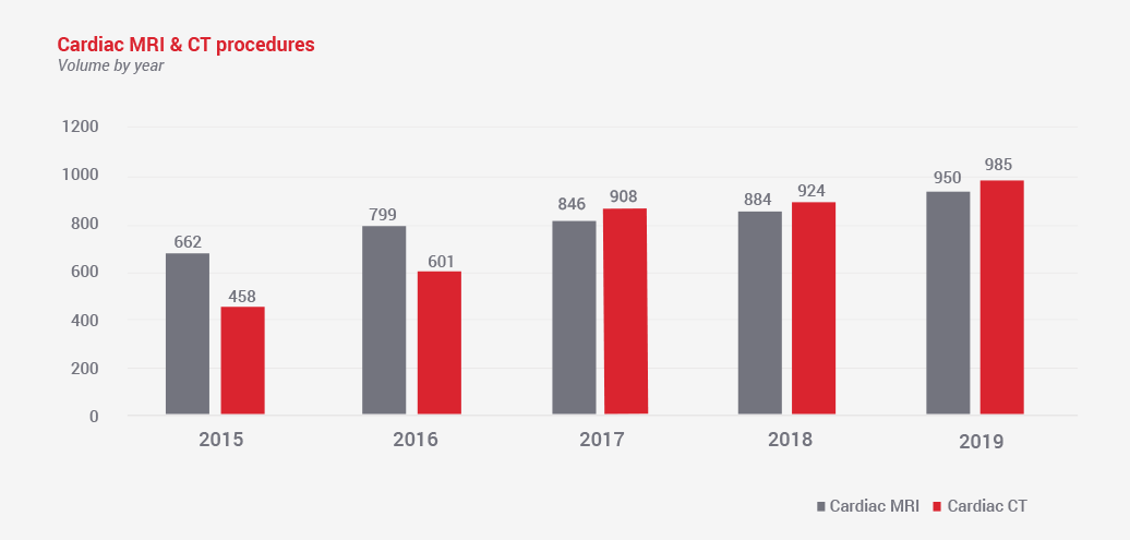
The diagnostic imaging capabilities of cardiac MRI and CT provide an excellent complement to the primary imaging modality, echocardiography. With advances in technology that allow rapid imaging with minimal radiation, cardiac CT has become vital in the diagnosis and surgical planning for many of our patients with congenital heart disease. For neonates with complex congenital heart disease, we use cardiac CT to provide rapid and excellent diagnostic information, typically without the need for sedation. For our single ventricle patients, we often employ cardiac CT prior to each surgical endeavor to supply information about the extracardiac anatomy to our surgeons.
Our cardiac MRI program is currently one of the largest in North America. This modality provides a wealth of information in multiple areas, including anatomy, morphology, volumetric and functional data, as well as physiologic information regarding blood flow and myocardial tissue characterization. Furthermore, cardiac MRI is frequently used in our teenage and adult patients with congenital heart disease because of the valuable information it provides and the limitations of transthoracic echocardiography among this population. In 2018, we opened our new hybrid cardiac MRI/interventional cardiac catheterization suite. This addition allows us to meld these two procedures together to combine the strengths of both modalities, decrease the use of radiation, and eventually perform MRI-guided cardiac catheterizations. Over the past year, we have already performed over 30 MRI/catheterization hybrid cases.
Pharmacologic Cardiac Stress MRI
At Texas Children’s Hospital, we are proud to have one of the busiest pediatric stress MRI programs in the world. There are numerous pediatric cardiac diseases that may be a cause of myocardial ischemia. Cardiac MRI is an excellent diagnostic tool to examine cardiac function, perfusion, and viability in these unique populations. As part of our robust Coronary Artery Anomalies Program, the majority of patients undergo a dobutamine stress MRI as part of their diagnostic work-up. For patients at risk of coronary ischemia, including those with an arterial switch operation or a diagnosis of Kawasaki disease, we are one of only a few pediatric centers in the U.S. to offer regadenoson as a pharmacologic stress agent. These tests require a dedicated, coordinated effort from a team of experts, including a pediatric cardiologist, pediatric radiologist, cardiac nurse, cardiac MRI technologist and MRI physicist. In 2019, our program performed over 150 cardiac stress MRI examinations.Advances in Cardiac Imaging
- Advanced methods to evaluate ventricular function by echo remain a research focus and are being integrated into clinical care. Cardiac strain analysis and 3D ejection fraction are being obtained in those who have single ventricle heart disease or have undergone chemotherapy or heart transplantation. Our research has helped determine that these technologies offer better reproducibility and accuracy with the ultimate goal of improving clinical care.
- Ventricular strain is also actively being investigated to assess for early ventricular dysfunction in other at risk patient populations, including those with chronic liver disorders or undergoing renal dialysis.
- 3D echocardiographic evaluation is offered to provide the most comprehensive imaging, which can be key to preoperative or pre-catheterization intervention planning.
- Quality improvement initiatives have been developed and enacted, so that we might deliver and preserve the highest quality of imaging for our patients.